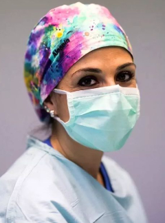
Fluorescence technology and its role in bariatric surgery
EAES Academy. Fassari A. 07/05/22; 366555; P298

Dr. Alessia Fassari
Contributions
Contributions
Abstract
Aim: Gastrojejunal and gastric leaks remain an important cause of morbidity and mortality after bariatric procedures (Laparoscopic Sleeve Gastrectomy, LSG; One Anastomosis Gastric Bypass, OAGB; Roux-en-Y gastric bypass, RYGB). Indocyanine Green Fluorescence (IGF) angiography is a recent development to assess tissue perfusion and perform vascular mapping during laparoscopic surgery which may help in preventing ischemia-related leaks.
Materials and Methods: As demonstrated in our video to perform LSG, almost all of the greater curvature blood supply is coagulated and divided, starting just a few centimeters from the pylorus and working cephalad until all the short gastric vessels near the left crus are coagulated and divided. The dissection is carried very close to the angle of His to avoid leaving behind a significant part of the fundus. Sleeve gastrectomy is then performed on a 36-Fr bougie using linear stapler. In the OAGB dissection should be started on the lesser curvature at the crow's foot in order to enter into the lesser sac by carefully freeing posterior adhesion between the stomach and pancreas. Once this is done, the first staple firing is done. The gastric pouch must be lengthy and narrow, measuring around 15-18 cm, with a 50-150 ml reservoir capacity. Unfortunately, anatomic studies have demonstrated that the vascular supply of the upper part of the gastric tube can be damaged during these procedures. Furthermore there are other potential causes of leaks besides ischemia, such as mechanical increase of intragastric pressure in the setting of decreased gastric volume. In certain cases, it may be a combination of both mechanical and ischemic events that leads to a leak. The OAGB is completed with antecolic gastrojejunostomy between the posterior wall of the gastric pouch and the antimesenteric border of the jejunum (approximately 150-200 cm distal to the ligament of Treitz). Once the stomach is resected (case 1) and gastrojejunal anastomosis is performed (case 2) 1,25 mg of indocyanine green solution is injected in a peripheral vein.
Results: A regular and homogeneous perfusion was observed along the entire gastric sleeve including the esophago-gastric junction (LSG) and the gastrojejunal anastomosis (OAGB). On the contrary, the excised specimen appeared devascularized at IGF imaging as expected. Operation time was 30 min for LSG and 40 min for OAGB with negative intraoperative and 2-day postoperative methylene blue test. Patients were discharged on postoperative day 2.
Conclusions: ICG fluorescence is widely used in multiple surgical specialities but has been introduced in bariatric only recently. Our video shows the safety and feasibility of using fluorescent angiography for gastric or gastrojejunal perfusion assessment.
{{ help_message }}
{{filter}}


